Your horse is lame, and you’ve narrowed down the problem to his hind end. If you’re like many horse owners, your next thought might be to check his hocks, maybe his fetlocks. But what about his stifles? For many, it’s the forgotten joint, hiding in the shadows of the numerous and often more-familiar hock issues.
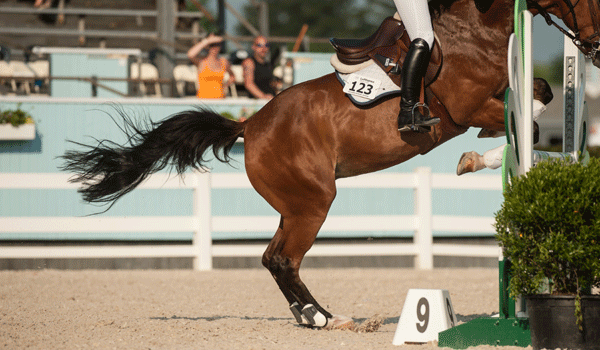
Yet the stifle is the largest, most complex system of joints in the horse’s body. It’s susceptible to a variety of joint disorders and cumulative wear and tear, as well as ligament and tendon injuries. And as diagnostics have advanced, veterinarians are discovering that this joint is the source of pain more often than previously realized.
With the help of David MacDonald, DVM, MVSc, DACVS, of Pioneer Equine Hospital in Oakdale, California, we’ll shed some light on the stifle’s anatomy, its function and common disorders to watch for in your horse.
As an Amazon Associate, Practical Horseman may earn an affiliate commission when you buy through links on our site. Product links are selected by Practical Horseman editors.
Anatomy and Function
The stifle is equivalent to the human knee, and like the knee, it’s comprised of four bones: the femur, the tibia, the patella and the nonfunctioning remnant fibula, which is fused to the tibia. These bones work together through three joints: the medial (inner) femorotibial joint, the lateral (outer) femorotibial joint and the femoropatellar joint (the joint between the femur and patella), which are all built to absorb shock and move your horse. Two cartilage-like discs called menisci act as a cushion between the femur and tibia, and 14 ligaments bind the joints together and help stabilize them by limiting range of motion.
The stifle’s job is to allow your horse to extend and flex his hind leg. To extend, the patella slips down over a bony knob on the lower section of the femur, the medial trochlea. When the horse brings his hind leg back underneath himself, the patella slips back up over the medial trochlea. The stifle also features the “passive-stay” mechanism, which allows the patella to essentiallylock in place over the medial trochlea, so the stifle remains extended and the horse can sleep standing up or otherwise bear weight on just one hind leg, allowing the other to completely relax.
Risk Factors and Trouble Signs
Sporthorses aren’t the most prone to stifle issues—that distinction likely goes to cutters and reiners, due to the nature of their work, with the abrupt stops and direction changes centered on their hind end, says Dr. MacDonald. But they are at greater risk than your average pleasure horse because of the added concussion, stress and strain placed on all of their joints and soft tissues, and the potential for injury—a slip, an impact with a jump or a kick.
Besides discipline, three additional factors can increase the likelihood of stifle trouble:
• Straight hind-leg conformation. The more upright the stifle angle, the less able it is to absorb shock, leading to increased concussion and stress—and thus increased wear and tear—on the joint.
• Poor muscle condition. Weak, lightly muscled stifles create a slackness that allows for too much movement in the joint, which again can add to the stress on the joint.
• Developmental disorders. These are problems that may be present when the horse is born or may develop as the foal grows, and include osteochondrosis and subchondral (under the cartilage) bone cysts, which will be discussed later in this story.
When stifle trouble strikes, the symptoms include heat, swelling and lameness as well as back and croup soreness, which are similar to those of hock problems, initially making diagnosis difficult, says Dr. MacDonald. In addition, the stifle-sore horse may exhibit common performance issues, such as stiffness, resistance to bending or picking up a particular lead, discomfort or unwillingness to go up or down hills, and drifting to one side when jumping.
Certain red flags are more likely to specifically indicate stifle trouble, including:
• difficulty picking up the correct lead behind
• trouble riding a transition from trot to canter or from canter to trot on a particular lead
• cantering in a “bunny hopping” manner
• dragging a hind toe
• a hitch or hesitation in the horse’s gait
“You’ll typically see the problem appear on the outside of the circle,” Dr. MacDonald notes. “That is, if the horse is exhibiting problems on the left lead, then think about the right stifle.”
Pinpointing the Problem
To definitively indicate the stifle as the problem center, your veterinarian will want to perform a thorough lameness exam, feeling for thickened ligaments, swelling, bone spurs and other palpable clues on the stifle, and then do flexion tests, in which he holds the limb flexed for 60 to 90 seconds and then your horse is immediately trotted off.
Once your vet is convinced the stifle is the point of pain, the exam probably will advance to nerve blocks, also called diagnostic intra-articular anesthesia. In this procedure, anesthesia is injected into a specific joint to desensitize the area surrounding it. If lameness is alleviated, the vet can more precisely identify the problem spot. “There are three joints in the stifle, so localizing the lameness is very important,” Dr. MacDonald says.
At this point, if your vet believes the stifle issue is fairly mild—there is not a lot of inflammation, for instance—he may recommend a conservative treatment plan, such as rest, nonsteroidal anti-inflammatories, intra-articular injections of an anti-inflammatory (such as hyaluronic acid and a corticosteroid) and a gradual return to work to rebuild condition. But because the horse often appears fine when first returned to work but then becomes sore again, your vet may recommend continuing down the diagnostic path to clarify the true underlying source of pain, Dr. MacDonald says.
In that case, the next step is likely to be radiographs to look for disturbances in the bone itself. If the X-rays look clean, then your vet might recommend an ultrasound to look for soft-tissue injuries. However, says Dr. MacDonald, “It’s not uncommon that radiographs and ultrasound don’t show anything abnormal. In a number of those horses, we’ll then use diagnostic arthroscopy,” in which a small arthroscope is inserted into small incisions in the stifle to view the joint. “In the last 10 years, this has become a valuable tool for diagnosing stifle lameness. We will sometimes see issues on the cartilage or even soft tissue that just don’t visualize any other way.”
Magnetic resonant imaging, often used to detect problems in other joints, usually isn’t a practical solution for investigating the stifle, says Dr. MacDonald. There are only a few MRI machines available that can accommodate the joint, and placement of the equine stifle in the magnet is limited by the horse’s size.
One Unique Disorder
Ultimately, the exam might show any of a number of issues. One that occurs only in the stifle is the intermittent upward fixation of the patella.
What it is:When a horse has IUFP, the stifle joint periodically locks in the extended position. This happens when the patella slides too far over that bony knob at the end of the femur, the medial trochlear ridge, and gets caught. Because the root cause can be slackness of the medial patellar ligament, “we talk about [the horse] having sloppy stifles,” says Dr. MacDonald.
How to spot it: Unlike many other stifle disorders, IUFP has a very distinctive symptom. It appears as a hitch in your horse’s gait—a hesitation between extension and flexion of the hind leg. The more severe the case, the longer the period of hesitation.
What causes it: Like other stifle problems, IUFP may be linked to straight hind-leg conformation, trauma or performance stress. It’s frequently tied to horses with poorly muscled quadriceps—the main muscles covering the stifle. (Even a normally fit horse who has lost condition due to time off may develop IUFP.) Other contributing factors may include a long toe–low heel hoof conformation, or deformations of the related bones and ligaments.
How it’s treated: There are multiple treatment approaches for horses with IUFP. The right one depends on the severity of the condition and the underlying cause. According to Dr. MacDonald, options typically include the following (roughly in order from least to most invasive):
• a conditioning or physical therapy program to strengthen the quadriceps
• nonsteroidal anti-inflammatory drugs (NSAIDs)
• corrective shoeing to elevate the heel
• blistering, which involves injecting a counter-irritant, such as a mixture of iodine and almond oil, into the affected area. “The thinking is that this scars the ligament,” says Dr. MacDonald. “That tightens the ligament down,” which helps keep the patella on track.
• medial patellar ligament splitting surgery, where the surgeon makes 10 to 15 small incisions in the ligament. Again, as the ligament heals and scars, it becomes thicker and tighter, and thus more stable.
• surgery known as a desmotomy, in which the surgeon completely severs the medial patellar ligament. More common 15 or so years ago, it’s still done in some cases that don’t respond to conservative treatment, says Dr. MacDonald. However, he adds, “the stifle has three patellar ligaments, and when you cut one, it changes how the patella tracks. And some horses will then develop arthritic changes at the end of the patella.”
• Estrone Sulfate treatment. “A dose of 10 mg is given as an intramuscular injection over a series of injections,” explain Dr. MacDonald. “This treatment has been very successful when combined with a quadriceps conditioning program.”
Of the many potential stifle disorders, IUFP is one of the few where treatment may yield an actual cure, rather than just management of the symptoms.
For your bookshelf: Kinesiology Taping for Horses: The Complete Guide to Taping for Equine Health, Fitness and Performance
Bone and Joint Disorders
Other common bone and joint disorders occur in various joints and include osteochondrosis, subchondral bone cysts and osteoarthritis. Of these, the first two are typically considered developmental disorders. The foal or young horse may display subtle symptoms, but trouble may not truly become apparent until the horse begins training. The last falls into the category of acquired disorders—those caused by things like trauma, and general wear and tear.
Osteochondrosis
What it is: Osteochondrosis essentially is a disorder where there is a failure of the cartilage template to form into bone (endochondral ossification), say Dr. MacDonald. When this happens at the joint surface, you can end up with bone fragments there. In the stifle, osteochondrosis is typically found on the femoropatellar joint and specifically on the lateral trochlear ridge, says Dr. MacDonald.
What causes it: Osteochondrosis is considered a multifactorial disease (multiple causes). It has been linked to nutrition—specifically, a young horse taking in too many calories or having an imbalanced calcium/phosphorous or zinc/copper intake—and to excessive strain on developing bones and joints. There may also be a genetic component, although that connection is still under debate.
How it’s treated: Treatment typically involves arthroscopic surgery to clean out the joint surface and remove the fragments, known as debriding the area.
Subchondral Bone Cysts
What they are: Cysts, also called lesions, are essentially cavities under the joint cartilage—gaps where bone should be, but isn’t. The cyst secretes inflammatory mediators, which spur a vicious cycle of inflammation, bone and tissue erosion, and, not surprisingly, pain. The cysts can grow larger over time, and the related inflammation can eventually lead to osteoarthritis.
In the stifle, bone cysts are most serious when they occur on the medial femoral condyle, which is a major weight-bearing point, says Dr. MacDonald.
What causes it: Subchondral bone cysts can be a manifestation of osteochondrosis and so share the underlying causes. In addition, traumatic injury may lead to the development of bone cysts.
Since this is a developmental disorder, the cysts are most common in horses age 3 and younger. They do, however, appear in older horses. In those cases, Dr. MacDonald speculates that the cysts may have existed since the horse’s younger years but didn’t present a problem until the horse’s protective articular cartilage eroded over time.
How it’s treated:There are a number of different ways to treat bone cysts, says Dr. MacDonald. “Traditionally, your veterinarian would inject the joint with an anti-inflammatory drug, such as hyaluronic acid and a corticosteroid, possibly complemented by an intra-muscular injection of polysulfated glycosaminoglycan (such as Adequan).”
For a horse who doesn’t respond to this approach, treatment may advanceto one of several options, including:
• surgical debridement of the cyst, although this tactic is not typically used today, since it presents a risk of the cyst enlarging after surgery, says Dr. MacDonald.
• injecting the cyst with the anti-inflammatory corticosteroid triamcinolone acetonide, using ultrasound and radiographic guidance. Veterinarians picked up this technique about 12 years ago from research in human medicine. “It was a revelation,” says Dr. MacDonald.
• a combination of the two, an approach based on research at Colorado State University around six to eight years ago. “We can go in with arthroscopy, look at the joint and where the cyst is, and we can debride any cartilage damage over the lesion, and then inject the cyst in multiple places with visual guidance. We’ve had success with that,” says Dr. MacDonald.
Osteoarthritis
What it is: As in other joints, OA in the stifle occurs when inflammation breaks down the cartilage in the joint and, eventually, causes the bone to produce more bone, causing pain and restricted movement. In its more advanced stages, OA can become degenerative joint disease.
What causes it:Torn cartilage, bone chips, displacement of a joint, subchondral bone cysts and the simple stress of daily performance demands can all cause or contribute to OA.
How it’s treated: There is no cure for OA, so treatment focuses on managing inflammation and pain. Depending on the degree of the problem, rest and NSAIDs might work. The next step up would be intra-articular injection of anti-inflammatory medications, says Dr. MacDonald, who also notes that IRAP can be useful as an anti-inflammatory.
Soft-Tissue Injuries
Like other ligaments throughout the horse’s body, those in the stifle joint—as well as the cartilage-like menisci—are susceptible to strains and tears. These result from trauma, and high-intensity sports like jumping and upper-level dressage can increase that risk.
Horses don’t often experience cranial and caudal cruciate ligament injuries, Dr. MacDonald notes, which would be equivalent to anterior cruciate ligament (ACL) tears in the human knee. When they do occur, he says, “They can be either minor, usually incidental to another issue. Or they can be really severe, and at that point, there is nothing you can do. You can’t replace it, and [without it], you lose the stability of [the joint].”
While ligaments and menisci can’t be replaced, modern treatment options, including stem cells and PRP, offer the possibility of regeneration and repair.
Regarding the long-term prognosis for stifle disorders, “You can’t make a blanket statement [about outcome],” says Dr. MacDonald. “It’s not the most forgiving joint,” and often the best you can do is treat symptoms rather than fix or cure the problem. The good news is that, even if all you can do is manage the disorder, today’s treatment options present more opportunity than ever for horses to overcome stifle issues and continue performing.
David MacDonald, DVM, MVSc, ACVS, has been a staff surgeon at Pioneer Equine Hospital in Oakdale, Calif., since 2000. His focus is surgery and lameness. Born and raised in Canada, he is a graduate of the Ontario Veterinary College-University of Guelph and the Western College of Veterinary Medicine. He became a diplomat of the American College of Veterinary Surgeons in 1997.
This article was originally published in the June 2014 issue of Practical Horseman.