
In equine veterinary medicine, computed tomography (CT) scans are primarily used to diagnose, evaluate and even guide treatment of leg and foot lameness as well as head-related issues, from skull fractures and bone infections to sinus and dental trouble. They can image both soft tissue and bone. Like the standard EEGs discussed in the preceding section, though, traditional CTs need horses to be immobile, which requires general anesthesia. In addition, the anesthetized horse has to be maneuvered into a scanning unit–and not every equine body part fits in a scanner, limiting its use.
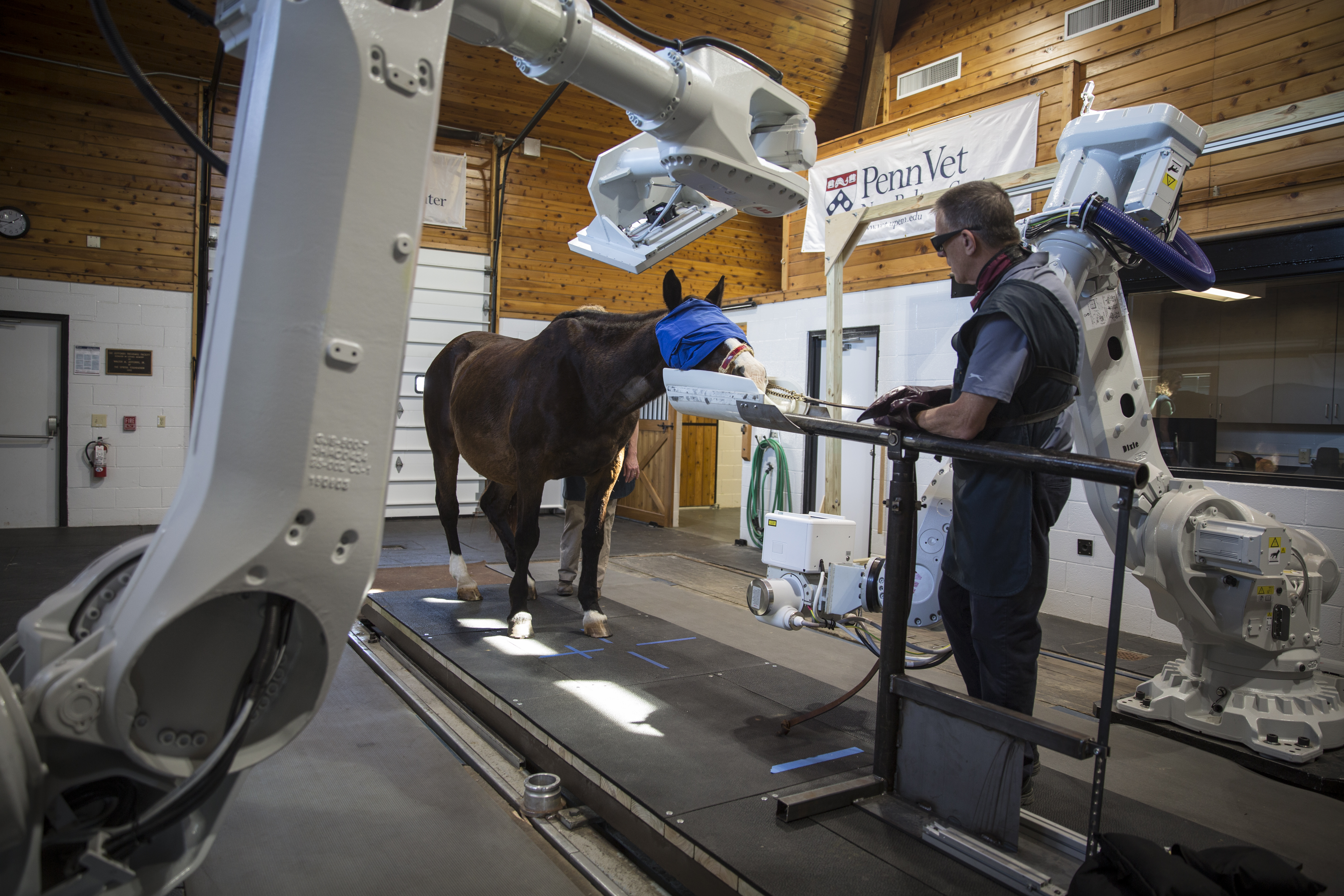
Now there’s a solution. The University of Pennsylvania’s New Bolton Center has become the world’s first veterinary teaching hospital to use a robotics-controlled CT scanner: the EQUIMAGINE system, developed by 4DDI with components from robot manufacturer ABB.
Since the system adjusts for some movement, the patient requires only sedation, not anesthesia, and can stand during the scan. During a procedure, the horse is led into position between two large robotic arms holding the scanning equipment. The arms move around the horse, collecting the necessary scans, which are reconstructed by a computer to produce high-quality, three-dimensional images.
In a video demonstrating the new system, Barbara Dallap Schaer, BS, VMD, associate professor of emergency medicine and critical care, explains that it provides much higher resolution and will allow veterinarians “to detect injuries at much earlier stages and prevent things that could be fatal either to the horse or, honestly, to the rider.”
The robotic CT scanner reduces risk as well as mental and physical stress for the horse and may cost less than a traditional CT. All of which could encourage more owners to take advantage of this valuable tool –particularly once the manufacturer begins filling its orders for more than a dozen additional units destined for equine facilities around the globe. One scanner has already been installed at the Cornell Ruffian Equine Specialists in Elmont, New York, and others are destined for equine hospitals in Kentucky, New Jersey, Texas and California.
As a bonus, the new CT system could well translate to the human world, says Dr. Schaer. For instance, she notes, “Instead of a child having to be anesthetized … they can sit there with their iPad and talk to their parents, and have the image performed in 30 seconds.”
This article was originally published in the August 2017 issue of Practical Horseman.