Run your hands down your horse’s legs and you’ll feel flesh and bones, tendons and ligaments. All are vital structures that work together to bear his weight and facilitate movement. But none play a bigger role than his joints in providing thrust and absorbing impact with every step he takes. Knowing a bit about their anatomy can help guide your efforts to preserve and maintain them to keep your horse moving soundly.

The Basics
A joint is formed where two or more bones connect. There are three types in the horse’s body: immobile fibrous joints (the sort that connect the bones of the skull), cartilaginous joints, which move only slightly (such as those found between the vertebrae of the spine) and synovial joints, which are the most common and most movable. They make up the majority of joints in a horse’s leg.
There are six different kinds of synovial joints, and their shape and construction determine how and to what extent they move. The two predominant types in a horse’s leg are
- ball-and-socket, where the rounded end of one bone fits into the cuplike end of another, allowing relatively free rotary motion (a great range of movement)
- hinge, in which two bones move in only one direction, back and forth, like the hinges on a door.
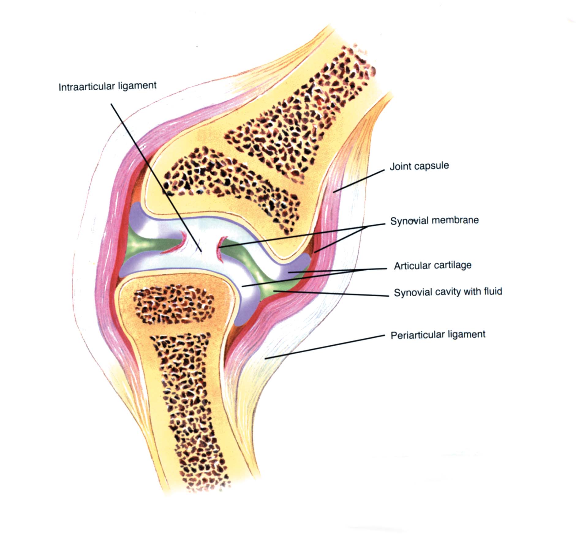
All synovial joints share the same basic structure: Each bone end, covered in smooth and resilient articular cartilage, is encased in a fibrous capsule that helps to provide stability. Collateral ligaments, made of very strong fibers, attach to the sides of the bones within the capsule. Other stabilizing ligaments and tendons, inside and outside the capsule, may also offer support during movement.
A synovial membrane lining the inner surface of the capsule secretes a clear, sticky fluid to lubricate, nourish and cleanse the joint. A chief component of the synovial fluid is hyaluronic acid, a viscous liquid that protects the surface of the articular cartilage during stress and impact. It also plays a role in combating potentially harmful inflammation. Changes in the normally protective quality of hyaluronic acid often are associated with joint disease.
How the Forelegs Stack Up
It’s estimated that a horse’s front limbs bear 60 to 65 percent of his weight. They also experience more force and concussion than the hind limbs, especially in horses that jump and race. As a result, the joints of the forelegs are more susceptible to injury and disease. They include:
shoulder—a highly mobile ball-and-socket joint that connects each front leg to the trunk and provides support for the front half of the body; formed by the scapula and humerus (upper arm bone)
elbow—a hinge joint of the humerus and the fused ulna and radius, allowing for flexion (bending) and extension
carpus (knee)—an extremely complex joint equivalent to the human wrist; made up of eight bones, organized in two rows, and three main joints: one between the radius and the first row of four carpal bones, one between the two rows of carpal bones and one between the lower row of carpal bones and the cannon bone
fetlock—a high-motion hinge joint that is the connection point for the cannon bone, proximal sesamoid bones and long pastern bone (first phalanx), supported by the suspensory ligament
pastern—a less mobile and less shock-absorbing hinge joint made up of the long pastern bone and short pastern bone (second phalanx)
coffin—a more mobile and usually highly shock-absorbing joint that connects the short pastern bone to the coffin bone (third phalanx).
The Hind Legs’ Joint Structure
Each hind limb of the horse runs from the pelvis to the navicular bone. The joints include:
hip—a ball-and-socket joint formed by the acetabulum and femur; allows the entire hind limb to swing back and forth and move sideways to swing outward and inward
stifle—the horse’s largest and most complex joint, equivalent to the human knee; made up of three bones—femur (thigh), tibia (shin) and patella (kneecap)—and three joints: two connecting the bones, one connecting the kneecap
tarsus (hock)—similar to the human ankle; links the tibia to the lower leg bones; made up of four joints formed by 10 bones and a number of stabilizing ligaments; the uppermost joint (tibiotarsal) is a ball-and-socket responsible for extensions and the majority of mobility; the lower three joints permit almost no movement and serve as shock absorbers; the lowest two of them are the ones typically affected by osteoarthritis
fetlock, pastern, coffin—refer to foreleg descriptions.
From Anatomy to Action
What can you do to protect and preserve the vital moving parts of your horse’s legs as well as the supporting structures? Consider the role played by his
- conformation. Shortcomings can lead to problems related to concussion—the force that travels up the leg each time a hoof hits the ground.
- weight. Extra pounds increase the load joints bear.
- hoof condition. Regular trimming to maintain balance helps hooves strike the ground evenly, minimizing stress on legs.
It’s also beneficial to
- use regular exercise tailored to your horse’s ability to maintain circulation to his joints and keep them mobile. Regular work consistent with a horse’s level of training also helps overall stability as well as muscle and core strength.
- take time—10 minutes of active walking is ideal— to warm up tissues and get joints working freely before introducing more strenuous work. Don’t over-drill during training. Do invest 10 minutes in cool down when your work is through.
- train and ride on good footing to reduce the possibility of missteps and lessen joint stress.
- perform a post-ride or daily leg check to detect heat, swelling and pain; study your horse’s movement to spot lameness. Consult your veterinarian if something looks or feels out of the ordinary. Detecting and treating joint degeneration (osteoarthritis) early on can slow its progression. Your veterinarian will recommend an appropriate course of care.
- feed wisely: Meet energy requirements with adequate protein and sufficient vitamins and minerals to maintain bone, cartilage and synovial fluid. Consult your veterinarian to determine your horse’s specific needs. Adding a nutraceutical supplement to his diet may be beneficial.
Practical Horseman thanks David Frisbie, DVM, PhD, DACVS, of the College of Veterinary Medicine and Biomedical Sciences at Colorado State University for his technical assistance in the preparation of this article. A professor in equine surgery in the Department of Clinical Sciences, Dr. Frisbie specializes in orthopedic research, equine lameness and using biologics, such as stem cell, to treat musculoskeletal injuries.
This article originally appeared in the Winter 2021 issue of Practical Horseman.


