You finish a weekend competition and your horse comes up just a bit off with some mild swelling and heat in his lower foreleg. After a few days off, he feels back to normal—and you feel like you dodged a bullet. But did you?
In reality, your horse may have started on the path to a serious tendon injury. If so, identifying and treating it promptly will be crucial to minimize the damage and maximize the chances of a full recovery. Here, Carol Gillis, DVM, PhD, DACVSMR, of Equine Ultrasound & Rehabilitation in Aiken, South Carolina, and Scott McClure, DVM, PhD, owner of Midwest Equine Surgery and Sport Medicine in Boone, Iowa, provide an overview of tendons, how injuries occur and what you can do to optimize the prognosis for your horse.
Q: What is a tendon?
A: Simply put, tendons are cables of soft tissue that connect muscle to bone. They largely consist of fibers or filaments made from the protein collagen. Living cells called fibroblasts create the fibers, forming an organized network of dense, elastic connective tissue.
Fibroblasts are also responsible for repairing damage to the collagen fibers. But since there are a relatively low number of fibroblasts compared to collagen, the need for repairs can sometimes outstrip the fibroblasts’ ability to catch up, which is one reason that tendons heal slowly.
A tendon’s primary jobs are to stretch and contract. In essence, they’re at work every time your horse moves. As your horse puts weight on a leg, for instance, the tendon stretches to take the load—picture a jumper’s front legs as he lands from a fence. The tendon contracts as the horse picks up the leg, removing the load—picture that same jumper’s forelegs as he’s in the air before a fence.
What tendons are most prone to equine injuries?
A: While tendons are associated with all muscles, there are two that often come into play with equine injuries:
The superficial digital flexor tendon can easily be seen behind the cannon bones. It attaches just above the elbow in forelimbs and just above the stifle, at the back of the femur, in hind limbs, ending at the long and short pastern bones. The SDFT acts on the knee, hock and fetlock joints.
The deep digital flexor tendon has three origination points in the forelimbs—the humerus, radius and ulna. It runs behind the knee and over the navicular bone, ending behind the coffin bone. In the hind limbs, the DDFT starts at two points on the tibia and ends behind the coffin bone. As its name implies, this tendon is placed deep beneath the SDFT. And like the SDFT, it’s involved with knee and hock action as well as forefoot, hind foot and elbow action.
Q: How do injuries occur?
A: Horses may experience an acute tendon injury, says Dr. McClure. This is where severe damage occurs from a one-time event. This is something like putting a foot wrong during a turn or on a jump landing. However, repetitive-use injuries are more common, he and Dr. Gillis agree. In these cases, “micro-tears accumulate over time until the tendon can no longer hold the load being imposed,” says Dr. Gillis, who notes that fatigue often plays a role.
These micro-tears may not initially cause your horse pain, particularly since tendons aren’t heavily loaded with pain receptors, says Dr. Gillis. Subtle signs may appear in the form of performance problems, which can easily be misinterpreted as behavioral or training issues, she adds. That can result in additional work—leading to additional damage—as the rider tries to correct the perceived problem.
“When enough fibers are damaged, clinical signs of pain, heat and swelling develop,” Dr. Gillis says. However, those symptoms may clear up quickly with some anti-inflammatory medication and rest—only to return when the horse starts back in work and fibers continue to tear.
Because of this vicious and potentially deceptive cycle, “many horses train and compete over months with tendon injuries that are increasing in severity,” Dr. Gillis says.
Q: What are some common types of tendon injury?
A: The most common tendon injuries, says Dr. McClure, are really just “a different degree of the same thing. A strain is just a lesser version of a bow. They have a very similar pathology.” Beyond such overstrain injuries, tendons can also suffer from edge tears, inflamed tendon sheaths, lacerations, infections, calcification, chronic scar tissue and compression from adjacent tissue, Dr. Gillis says.
The DDFT, in particular, can incur damage where it wraps around the navicular bone. While this often occurs in conjunction with degeneration of the bone, Dr. McClure says it’s not always apparent which came first—bone or tendon damage.
Q: Are certain disciplines more prone to tendon injury than others?
A: “Horses’ tendons and ligaments have a limit as to how far they can stretch before fiber tearing occurs,” Dr. Gillis says. “Tendons are near their breaking strength at a gallop, over fences and in other full athletic endeavors.” Faster speeds, higher jumps, tighter turns and uneven ground all add strain. Not surprisingly, that puts nearly any performance horse at risk.
Dressage horses are prone to hind-suspensory-ligament and back injuries, Dr. Gillis says. Jumpers, adds Dr. McClure, are more prone to inflammation in the tendon sheath in their rear legs, while event horses may see more SDFT bows.
Q: How can riders prevent tendon injuries?
A: The best way to prevent tendon trouble is to start with a horse who has good conformation. Drs. Gillis and McClure note that the following faults increase stress, which can put a horse at greater risk:
- Back at the knees
- Straight hind limbs
- Long back and short croup
Other factors that can strongly influence risk include:
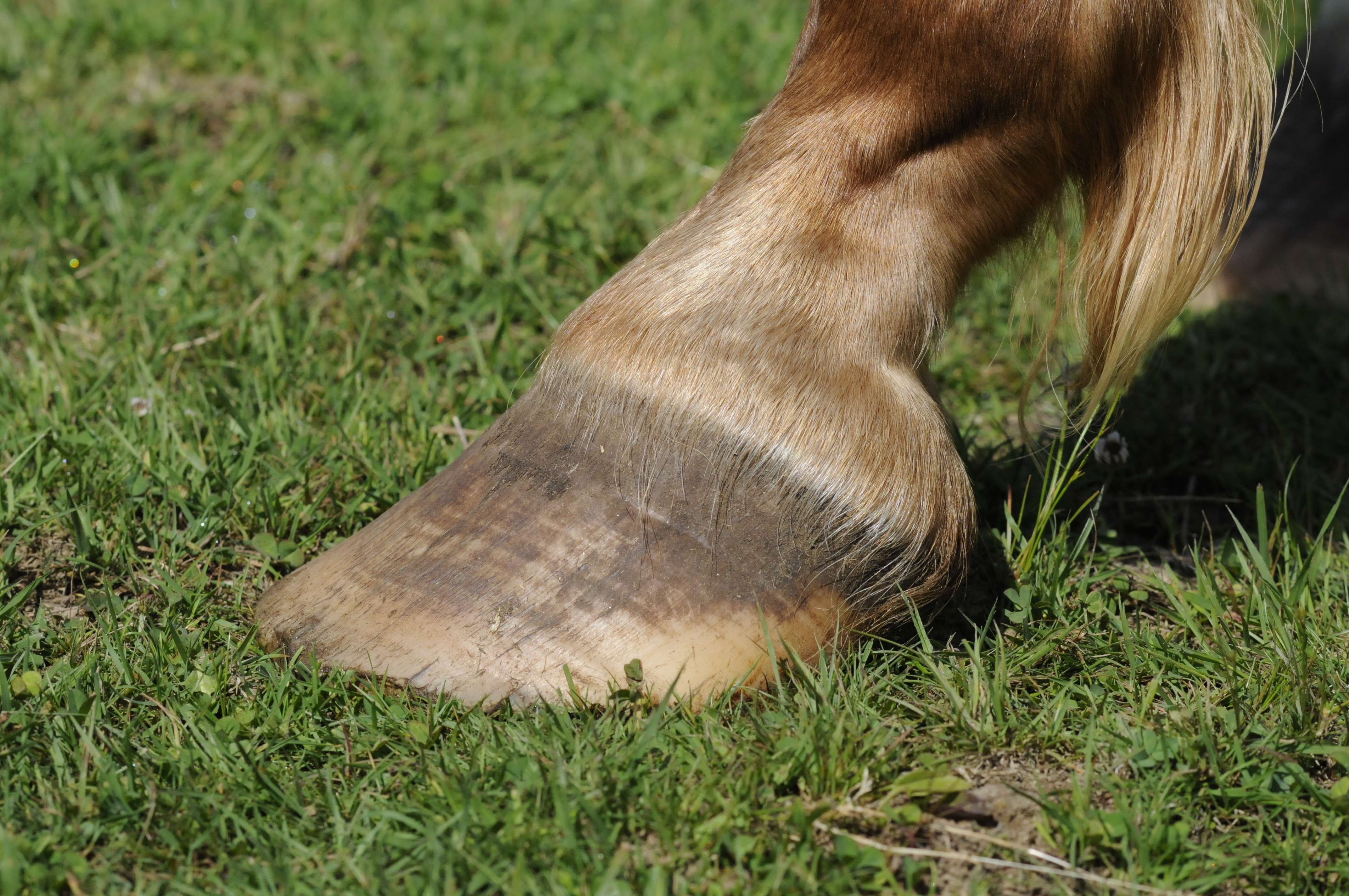
Hoof conformation and farrier work
Long toes, low, underslung heels and uneven heel heights can lead to injury-inducing strains. Larger grabs and caulks can also increase stress, Dr. McClure says.
Behavior
Horses who kick or bang their stalls or with a propensity for getting cast are at increased risk, so it’s important to address these issues as part of prevention, Dr. Gillis says.
Other body pain
Horses with sore feet, hocks or backs may compensate for the discomfort by placing extra weight on their tendons, thus increasing the chance of injury. Make sure that poor-fitting or improperly used tack isn’t contributing to overall soreness.
Footing and terrain
There’s some debate regarding what footing is worse on soft tissues and what risk hill work may pose. Dr. McClure notes that racehorses tend to have more tendon injuries when running on softer, deeper tracks. And, he says, Japanese racehorse trainers are among those who routinely train and condition on hills, and he doesn’t believe that hill work is a risk factor. Dr. Gillis counters that horses evolved as plains animals and that hill work has been proven to increase strain.
The main idea to remember, says Dr. McClure, is that “all of these things contribute a small piece.” But if there is one absolute must-have for your tendon-health toolbox, it’s a solid foundation of fitness, he adds. Not only can this help maintain your horse at an appropriate weight (itself an aid in preventing strain-related injuries), but proper conditioning also promotes soft-tissue growth and repair, notes Dr. Gillis.
She recommends that horses have ridden exercise four to five days per week with sport-specific exercise one to two days per week to prepare for competition. Fitness should be built gradually, with increases of about 5 percent per week to gain condition while mitigating risk. Fifteen-minute walk warm-ups are important to stretch the tendons so that significantly higher forces may be required to damage the tissue.
Q: How are tendon injuries diagnosed?
A: In the case of an acute tendon injury caused by a one-time event, Dr. McClure says, you’ll see immediate heat, swelling and lameness. But with a repetitive-use injury, which occurs over time, initial signs can be subtle. A little heat or swelling might be evident after exercise. Slight, intermittent lameness may appear a few days after a hard workout and then seem to resolve with rest or treatment. Poor performance can be another hint of trouble as can a less-than-expected response to a joint injection.
Since damage can be cumulative, an early diagnosis gives your horse the best chance at a shorter recovery period and a better long-term prognosis. So it’s best to have your veterinarian complete an exam before symptoms become severe, although that doesn’t mean setting an emergency appointment. In fact, a little delay may act in your favor as lesions can become more readily visible once any swelling has subsided.
For instance, says Dr. McClure, if you compete your horse over the weekend and see a little swelling Monday, have the vet examine it promptly. Then schedule a recheck in seven to 10 days, by which time the true extent of the injury should be evident.
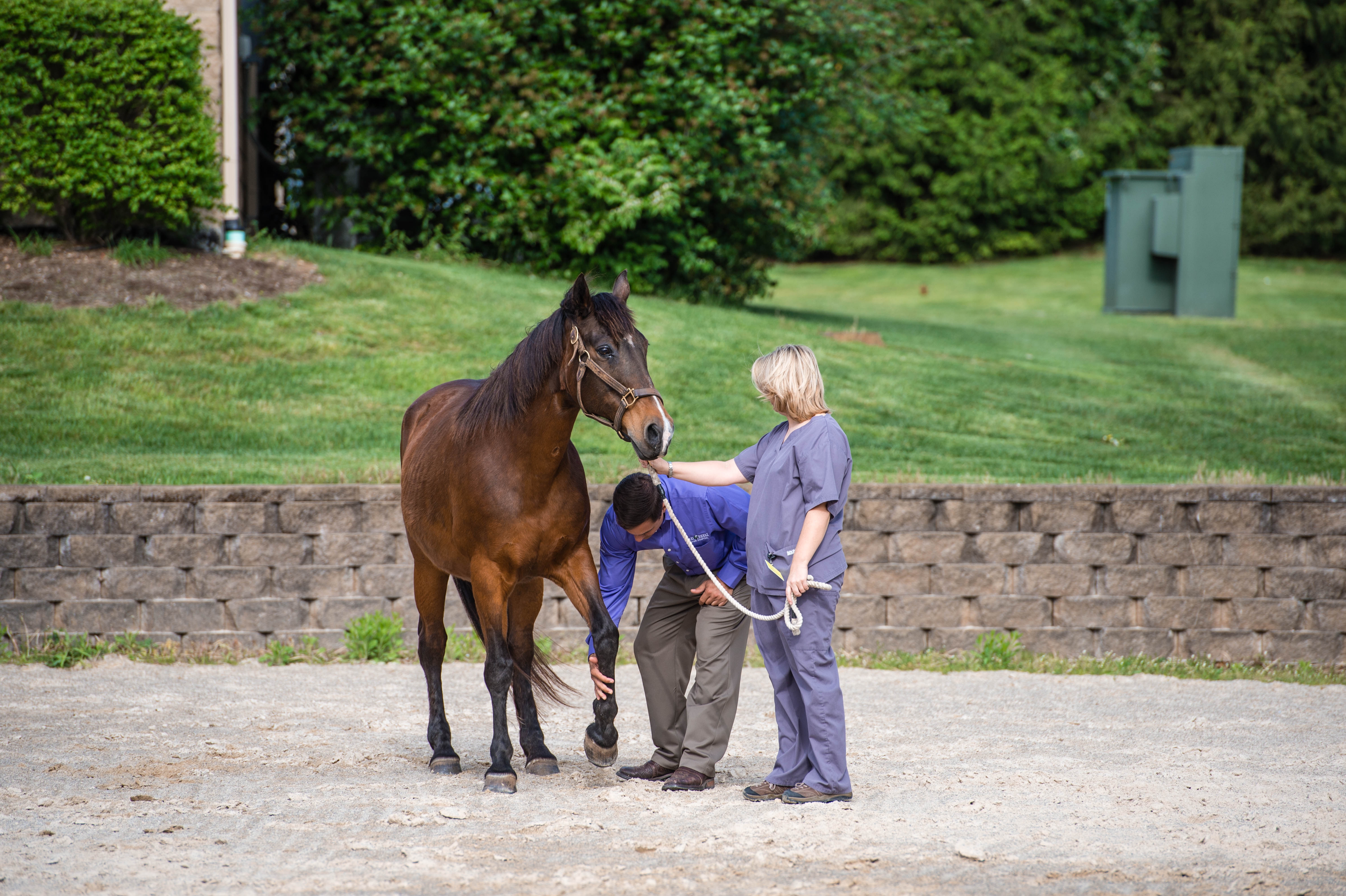
The exam itself should start with a thorough lameness evaluation, including palpation, watching your horse in action, stress or flexion tests
and possibly diagnostic nerve blocks to more precisely define the injured area, Dr. Gillis says.
Once the veterinarian has zeroed in on the most likely area of injury, ultrasound is typically the diagnostic tool of choice. “Radiographs, nuclear scintigraphy, CT [computerized tomography] and MRI [magnetic resonance imaging] can also provide information about the relationship of tendon injury to bone and cartilage,” she adds.
After the veterinarian has defined the injury, you should also discuss factors that might be contributing to tendon overload which, if uncorrected, could cause injury recurrence on return to work, Dr. Gillis says.
Q: How do you treat tendon injuries?
A: “The starting point [for treatment] is always the same,” Dr. McClure says. “Get the initial inflammation under control.” This step is critical because while inflammation is part of the healing process, it can also worsen tissue damage.
Generally, your veterinarian will likely recommend the following steps to modulate inflammation:
- Cold-water hosing or time standing in ice water. You might also try a high-tech cooling system. Twice-daily sessions should last at least 20 to 30 minutes.
- Wrapping with a thick quilt and track bandage to reduce swelling. Wrap the opposing leg, too, to manage edema in case the horse loads it more in an attempt to keep weight off the injury. Redo the wraps daily.
- Medicating with a nonsteroidal anti-inflammatory agent, such as phenylbutazone (bute), flunixin meglumine (Banamine) or diclofenac (as in the topical cream Surpass), which not only assists with swelling but also helps manage pain.
- Stall confinement. However, Dr. Gills recommends hand-walking, too, except in less-common cases where the injury is severe enough that rupture is likely or the horse is lame at a walk. Short, twice-daily sessions can help prevent joint stiffness, promote tendon gliding and deter adhesion formation. (Adhesions are essentially strands of scar tissue.) Adjust your horse’s diet for the reduced exercise and head off potential boredom (and accompanying bad behavior) by supplying plenty of good quality hay plus a pal in sight, Dr. Gillis says.
The cool-down process can take anywhere from a couple of days for a mild problem to as much as four weeks for a severe injury. As you feel the heat and swelling dissipate, call your veterinarian to schedule a follow-up ultrasound. Without the swelling, says Dr. McClure, you’ll have a good clean vision of the injury—how big it is, where it’s located—and those factors will affect the rehabilitation program and long-term prognosis.
At this juncture, your veterinarian may also discuss options for ancillary treatments, such as stem cells or shockwave therapy (see sidebar, pg. 65). Whether or not you decide to pursue one of these options may depend on the injury itself as well as your horse’s age, discipline and, of course, your budget, Dr. McClure says.
Q: After the cool-down phase, how should the recovery plan progress?
A: Your veterinarian should perform ultrasound re-evaluations every six to eight weeks to assess healing, Dr. Gillis says. The results will also provide a guide for how to proceed with each successive step of rehab.
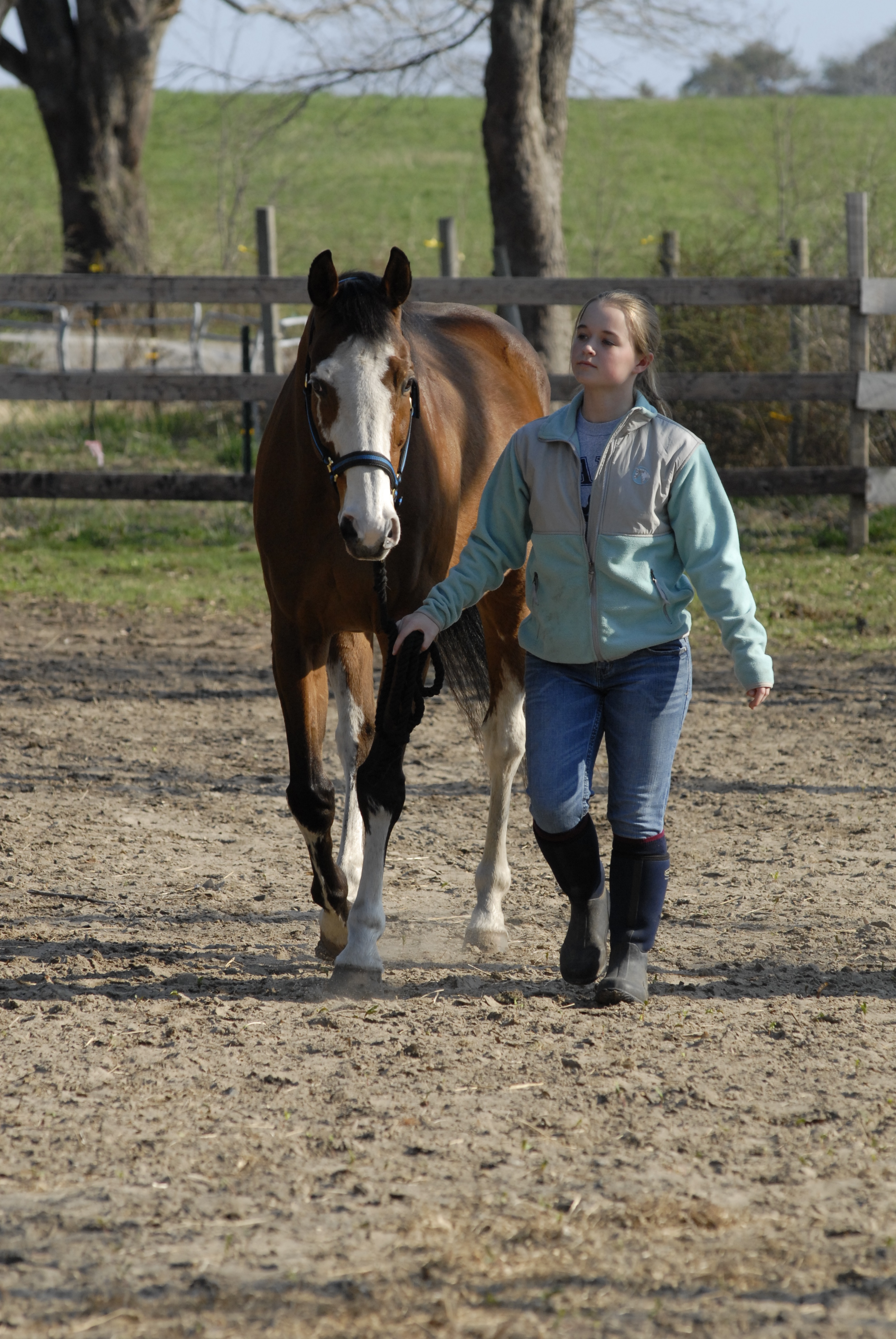
As a general rule, rehab will center on a program of controlled, graduated exercise, Dr. McClure says. Specifically, Dr. Gillis describes the following as a typical progression for most tendon injuries: two months of hand-walking; two months of walking under saddle; two months of walk and trot under saddle; and, finally, two months of walk, trot and canter under saddle. Only then is the horse likely ready to return to training for his specific sport with another six to eight weeks of training before returning to competition.
“More severe injuries require a longer healing time, while mild injuries may progress at six-week rather than eight-week intervals,” Dr. Gillis says. “This is where vigilance and early detection
pay off.”
The point of a graduated exercise program isn’t simply to get your horse’s muscles and cardiovascular system back in shape. By lightly loading the tendon, you actually encourage collagen formation. Over time, as strong, organized networks of collagen begin to repair the injury, the tendon gets stronger and can handle additional load.
But be cautious and patient: If you ask too much too soon, you risk re-injury. That’s why your veterinarian might initially recommend increasing work by, say, five minutes every two weeks. In addition, keep your work on level, smooth footing and work in straight lines with some large circles.
Throughout the early weeks of rehab, you may want to continue confinement to prevent your horse from overdoing it and reinjuring himself during turnout. When your veterinarian says turnout is OK, put him in a small private paddock, if possible. Make sure to supervise him. He may need a mild sedative if he’s having trouble staying calm.
Your veterinarian likely will recommend keeping your horse in wraps for several weeks after the cool-down phase, then gradually reduce the time spent in wraps.
Q: What are common setbacks during recovery?
A: Increasing workload before your horse is ready is the most common cause of a setback in healing. That’s where ultrasound follow-ups and your veterinarian’s experience are critical in guiding each step of your horse’s rehab.
Adhesions are another common bump on the rehab road. They can restrict the tendon’s ability to stretch and glide and can make your horse sore. However, the best therapy for this particular problem is to continue with your exercise plan. Movement will help to remodel the adhesions so that they no longer cause problems.
You can’t tell from the outside whether overwork or adhesions cause new soreness, heat or swelling. So consult with your veterinarian before determining whether to reduce your horse’s workload or press ahead.
Q: What’s the prognosis for a tendon injury?
A: Recovery from anything but the mildest tendon injury can take from nine to 12 months. A severe tear will take longer to heal than a moderate strain, and an older horse will probably heal more slowly than a younger one.
Placement of injury and the horse’s discipline matter, too. For instance, says Dr. McClure, jumpers with a hind-limb DDFT injury tend to have repeat issues. A horse sound with an injury where the DDFT attaches to the navicular bone can be hard to keep sound.
One reason horses may not return to prior form—fibers in a repaired tendon can be less elastic. This makes them more prone to strains and tears than the collagen fibers of a normal, healthy tendon. But a good rehab program can positively influence the development of more normal, healthy collagen fiber, agree Drs. McClure and Gillis—and that can mean a huge difference for a horse’s long-term prognosis.
“In my practice, 85 percent of horses which successfully complete a full rehab program return to their intended athletic use. They are in the same risk category as a previously uninjured horse,” Dr. Gillis says. “Of the 15 percent of horses that heal the soft-tissue injury but do not return to the previous level of athletic endeavor, the majority have bone or joint issues that cannot be successfully treated.”
Therefore, Dr. McClure says that giving your horse his best chance of a full recovery is about the whole program. It’s not about just one step in the process. Your patience, attention to detail and commitment to following the entire program correctly can pay off in a big way. The hopeful end result: You back in the saddle and your horse back in top form.

Ancillary Treatments
When it comes to treating soft-tissue injuries, researchers continue to seek methods that will promote healthy, normal collagen-fiber growth. They also want to diminish the development of scar tissue and maximize the odds of full recovery. Below are some of the options you may want to discuss with your veterinarian. However, Dr. Gillis cautions that these treatments require further research to determine their most effective use.
Stem Cells
Stem cells can transform into different types of tissue—in a damaged tendon, they become fibroblasts, which make collagen. They can also produce growth factors that stimulate healing. A veterinarian can collect stem cells from your horse’s fatty tissue or bone marrow. She then concentrates and purifies it before injecting it into the injury site. A new technique for culturing stem cells is being studied at the Animal Health Trust in Newmarket, England. It involves adding growth factor to the stem-cell culture and growing the cells on a “scaffold.” The goal is to produce more of the collagen.
Extracorporeal Shock-Wave Therapy
Extracorporeal shock-wave therapy uses high-energy pressure waves to improve blood flow, stimulate growth factors and reduce inflammation. Dr. McClure says it may not speed the rate of healing. But it may repair the tendon with a little better quality tissue.
Platelet-Rich Plasma
Platelet-rich plasma is another common ancillary treatment. Platelets circulate naturally in the blood and contain substances that promote healing, stimulate blood- vessel formation and control inflammation. Veterinarians can separate platelets from a sample of the injured horse’s blood. Then they can process it so there is a concentrated dose in the plasma (the liquid portion of blood). They then can inject the PRP into the injured areas. They often use it in combination with stem cells.
Interleukin-1 receptor antagonist protein, also known as autologous conditioned serum, is derived from your horse’s own blood. Interleukin-1 is an inflammatory mediator that can exacerbate a tendon injury. IRAP processes the blood sample to create a plasma rich in a protein that prevents IL-1 from binding to its receptors. This effectively blocks it from causing added inflammation.
Extra Cellular Matrix
Extra cellular matrix, unlike the preceding treatments, doesn’t use live cells to aid healing. Instead, ECM, marketed as ACell Vet, is a scaffold of protein fibers. Scientists formulate it as a powder that’s suspended in saline solution and then injected into the injury site.
Hyperbaric Oxygen Therapy
Hyperbaric oxygen therapy places the injured horse in an airtight, stall-like chamber at increased atmospheric pressure. The goal is to deliver a much higher level of pure oxygen. Research indicates that this may improve tissue healing and shorten recovery time. It does this by helping to reduce swelling, increase cell division and decrease oxidation.
Swimming
Swimming or an aquatreadmill can help your horse gain cardiovascular and muscular fitness with less strain on the tendons. Horse-care managers often use it to supplement under-saddle exercise.
Experts consider other treatments as experimental or unproven. These include therapeutic ultrasound and low-level laser therapy. They also include the injection of proteins or use of gene therapy to introduce growth factors to the injury site.
Practical Horseman published this article in the October 2016 print issue.


